Echo Normal Values Pdf
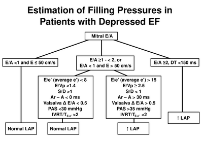
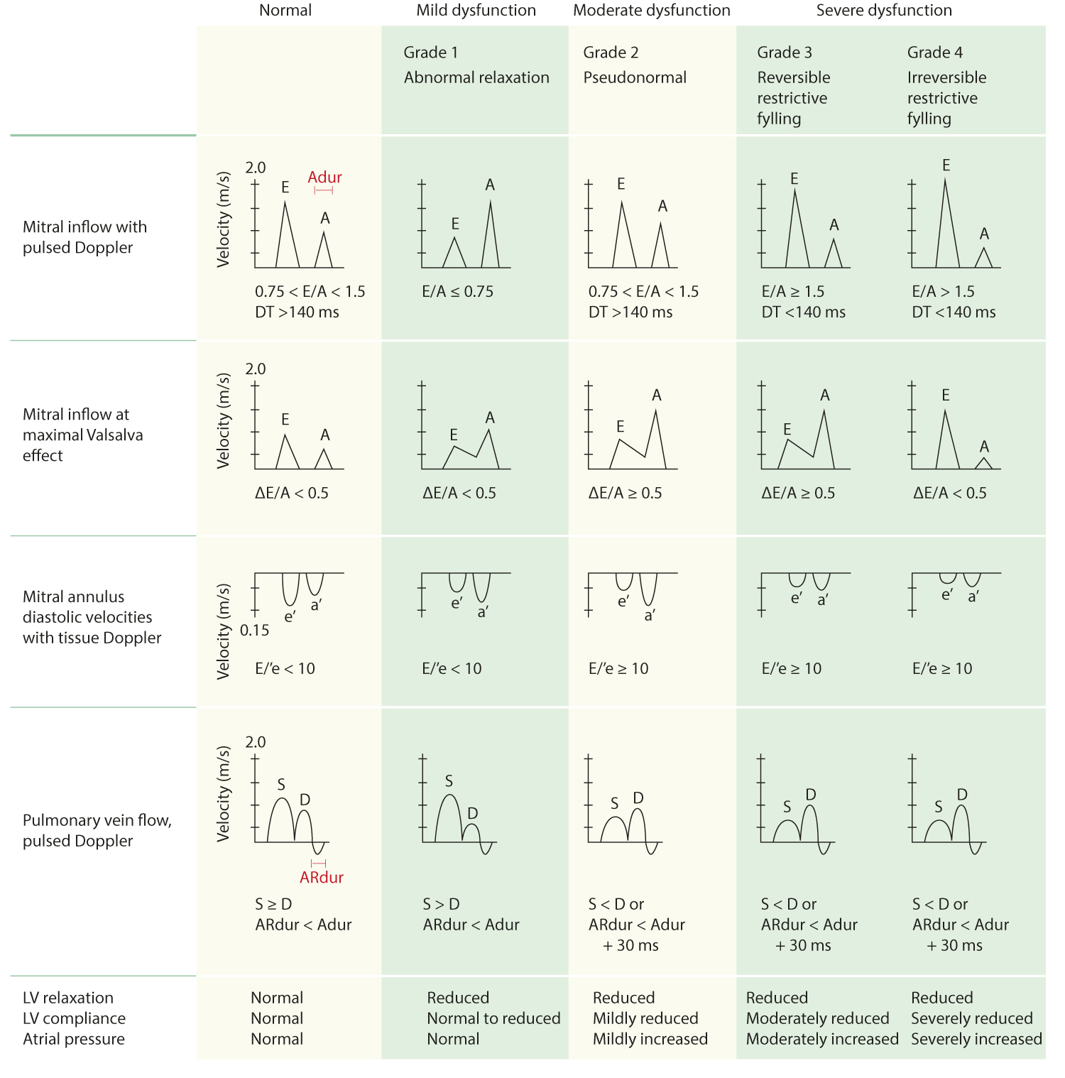
Tr velocity ≤ 2.8 m/sec 4. The right atrium is normal.


Aims availability of normative reference values for cardiac.


Echo normal values pdf. A total of 1152 clinically normal adult dogs, ranging in size from 1.4 to 97.7 kg, are represented (table 2). A routine transthoracic echocardiogram was performed. E/e’ ≤ 14 (ave), 15(med) 3.
Official publication of the american society of echocardiography 30 (4), pp. Reference ranges for different anatomical levels using different (i) measurement conventions and (ii) at different times of the cardiac cycle (i.e. The left ventricular mass of the white participants was 121.5±34.4g and in black participants was 124.2±33.6g (p=0.77).
In journal of the american society of echocardiography : The spleen (s) is more echogenic (hyperechoic) than the liver (li), which is the same or slightly more echogenic (brighter) than the cortex of the kidney (ck). Asch, fase, jose banchs, fase, rhonda price, vera rigolin, fase, james d.
Echocardiography (echo).2,3 the normal pericardium consists of two layers: Thus to normalize in pediatric echocardiography we use nomograms normalized data are expressed as z score i.e. R normal echogenicity rule of thumb:
The dimension of the right ventricle was similar in both sexes. A z score of +2 or 2 corresponds to the 95th percentile (i.e., 2 standard deviations above or below the mean) nomograms and z scores. A z score > 2 indicate dilatation while a z score <2 indicate hypoplasia.
A report from the american society of echocardiography developed in collaboration with the society for cardiovascular magnetic resonance. Normal values for echocardiographic measurements ficantly higher in men. Eroa = (csa*v)/v (maxmr) { { flödeshastighet = csa*v = 6.28*r2*v (aliasing) references.
The 5th and 95th percentiles of the echocardiographic parameters are presented in table iii. Based on results from this study we proposed normal reference values for the acquired echocardiographic measurements using the mean ± 2 standard deviation rule, which is based on the assumption that 95% of values of a reference group fall into this range, in such a way that 2.5% of the time a sample value will be less than the lower limit of. The right ventricle is normal in size with.
∗∗in the absence of other etiologies of lv and la dilatation and acute mr. The left atrium is normal. The mitral valve is normal.
The visceral pericardium, which is contiguous with the epicardial surface of the heart, and the parietal pericardium, which is a thin fibrous structure closely apposed to pleural surfaces laterally and that merges with the diaphragm inferiorly. The aortic valve is normal and trileaflet. Establishing normal echocardiographic values editorial comment need for a global definition of normative echo values—rationale and design of the world alliance of societies of echocardiography normal values study (wase) federico m.
E’ velocity ≥ 7(med), 10 (lat) cm/s 2. Conclusion the norre study provides normal values of proximal aorta dimensions as assessed by echocardiography.


0 Response to "Echo Normal Values Pdf"
Post a Comment